DESCRIPTION
AMMONIA (NH3) or its ion form ammonium (NH4 + ) is an important source of nitrogen for living systems. It is synthesized through amino acid metabolism and is toxic when present at high concentrations. In the liver, ammonia is converted to urea through the urea cycle. Elevated levels of ammonia in the blood (hyperammonemia) have been found in liver dysfunction (cirrhosis), while hypoammonemia has been associated with defects in the urea cycle enzymes (e.g. ornithine transcarbamylase).
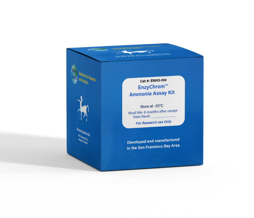
Simple, direct and automation-ready procedures for measuring NH3 are popular in research and drug discovery. BioAssay Systems' ammonia assay is designed to directly measure NH3 and NH4 + . In this assay, NADH is converted to NAD+ in the presence of NH3, ketoglutarate and glutamate dehydrogenase. The decrease in optical density at 340 nm or fluorescence intensity at λem/ex = 450/360 nm is directly proportionate to the NH3 concentration in the sample.
KEY FEATURES
High sensitivity and wide linear range. Use 20 µL sample. Linear detection range 24 to 1000 µM ammonia.
Homogeneous and simple procedure. Simple “mix-and-measure” procedure allows reliable quantitation of NH3 within 30 minutes.
APPLICATIONS
Direct Assays: NH3 in biological samples (e.g. serum, plasma, urine, saliva, cell culture etc).
KIT CONTENTS (100 TESTS IN 96-WELL PLATES)
Assay Buffer: 20 mL Enzyme: 120 µL
Ketoglutarate: 120 µL Standard: 400 µL
NADH Reagent: Dried
Storage conditions. The kit is shipped on ice. Store all components at -20°C. Shelf life of six months after receipt, 3 weeks after reconstitution.
Precautions: reagents are for research use only. Normal precautions for laboratory reagents should be exercised while using the reagents. Please refer to Material Safety Data Sheet for detailed information.
PROCEDURES
Reagent Preparation. Equilibrate all components to room temperature. Briefly centrifuge all tubes before opening. Reconstitute the NADH Reagent tube with 1000 µL dH2O (final 10 mM). Unused reconstituted NADH reagent is stable for three weeks when stored frozen at -20°C.
Sample preparation: solid samples can be extracted by homogenization in distilled water (dH2O) and filtered, centrifuged or, if necessary, deproteinized to remove any undissolved material. Samples should be clear and colorless with pH adjusted to 7 - 8. Serum and plasma samples can be assayed directly. Cell culture media should be diluted 5-10 fold in dH2O prior to assay.
Colorimetric Procedure
1. Standards and Samples. Prepare a 1000 µM NH3 Standard Premix by mixing 15 µL of the 20 mM Standard and 285 µL dH2O. Dilute Standard as follows.
Transfer 20 µL standards into separate wells of a clear, flat-bottom 96-well plate.
Transfer 20 µL of each sample into two separate wells, one serving as a sample blank well (RBLANK) and one as a sample well (RSAMPLE).
2. Enzyme Reaction. For each standard and sample well, prepare Working Reagent by mixing 180 µL Assay Buffer, 1 µL Enzyme, 8 µL reconstituted NADH Reagent and 1 µL Ketoglutarate. Add 180 µL Working Reagent to the four Standards and the Sample Wells.
Prepare blank control reagent by mixing 180 µL Assay Buffer, 8 µL reconstituted NADH Reagent and 1 µL Ketoglutarate (No Enzyme). Add 180 µL Blank control reagent only to the Sample Blank Wells. Tap plate to mix. Incubate 30 min at room temperature.
3. Read OD340nm. Fluorimetric Procedure The fluorimetric procedure is the same as for the colorimetric assay, except that a black, flat-bottom 96-well plate is used. After incubation for 30 min at room temperature, read fluorescence intensity at λex = 350-360 nm and λem = 450 nm. CALCULATION Subtract the standard values from the blank value (#4) and plot the ∆OD or ∆F against standard concentrations. Determine the slope and calculate the NH3 concentration of Sample,
RSAMPLE and RBLANK are optical density or fluorescence intensity readings of the Sample and Sample Blank, respectively. n is the sample dilution factor. Note: if the calculated NH3 concentration is higher than 1000 µM, dilute sample in dH2O and repeat assay. Multiply result by the dilution factor n. Conversions: 1000 µM NH3 equals 1.7 mg/dL or 17 ppm.
MATERIALS REQUIRED, BUT NOT PROVIDED
Pipeting devices, and clear flat-bottom 96-well plates and optical density plate reader for colorimetric assays; black flat-bottom 96-well plate and fluorescence intensity plate reader for fluorimetric assays.
PUBLICATIONS
1. Jiang, S., Yan, W., Wang, S. E., & Baltimore, D. (2018). Let-7 suppresses B cell activation through restricting the availability of necessary nutrients. Cell metabolism, 27(2), 393-403.
2. Zhu, J., Zhu, X. G., Ying, S. H., & Feng, M. G. (2017). Effect of vacuolar ATPase subunit H (VmaH) on cellular pH, asexual cycle, stress tolerance and virulence in Beauveria bassiana. Fungal genetics and biology, 98, 52-60.
3. Ono, M., & Yoshiga, T. (2019). Cellular immunity in the insect
Galleria mellonella against insect non-parasitic nematodes.
Parasitology, 146(6), 708-715.
